|
Chapter 3 The Different Parts of the Nervous
System 75
|
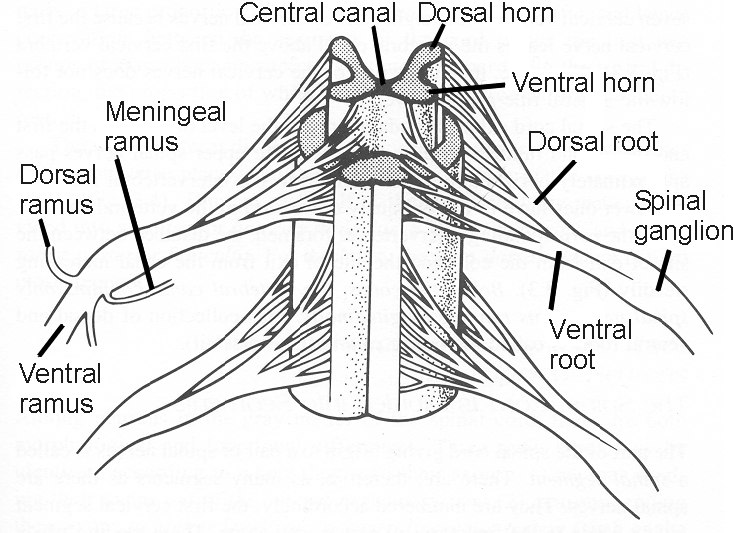
| Figure 3.5 Two segments of the spinal cord (seen
from the ventral aspect). In
the upper part, the white matter has been removed.
The dorsal and ventral roots emerge from the posterior and
anterior lateral sulci, respectively, and unite to form spinal
nerves. Note the location of the spinal ganglion at the site
where the roots unite. |
lies in the intervertebral foramen just where the dorsal
and ventral roots unite (Fig. 3.6). There is an important functional
difference between the ventral and dorsal roots: The ventral
roots consist of efferent (motor) fibers, and the dorsal roots of
afferent (sensory) fibers.
In total, 31 spinal nerves are present on each side, forming
symmetrical pairs (Fig. 3.3). They all leave the vertebral canal
through the intervertebral foramina on each side. As mentioned, the
ventral and dorsal roots unite at the level of the intervertebral
foramen to form the spinal nerves. The spinal nerves are numbered
(as a general rule) in accordance with the number of vertebrae above
the nerve. We therefore have 12 pairs of thoracic nerves, five pairs
of lumbar nerves, and flve pairs of sacral nerves. In humans, there
is only one pair of coccygeal nerves. There are
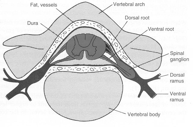 |
| Figure 3.6 Cross section of the vertebral column
showing the positions of the spinal cord and the spinal
nerves. Note the location of the spinal ganglion in the
intervertebral foramen. The spinal cord is surrounded by the
cerebrospinal fluid contained within the dura. Outside the
dura, there is fat and avenous plexus, also serving as soft
padding for the cord and the spinal nerves.
|
=> page 76
start page
|