|
Chapter 3 The Different Parts of the Nervous System
73
74 MAIN FEATURES OF STRUCTURE AND FUNCTION
 |
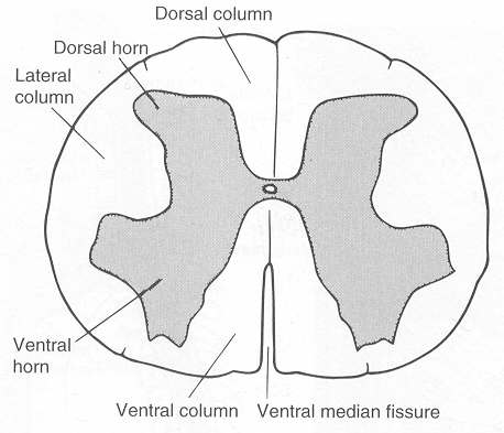
| Figure 3.4 Cross section of the spinal cord at a
lumbar level. Note the central H-shaped region of gray
matter and the subdivision of the surrounding white matter
into funiculi. |
column growing more rapidly than the spinal cord. In early
embryonic life, the neural tube and the primordium of the vertebral
column are equally long (Fig. 3.12).
The spinal cord is somewhat flattened in the anteroposterior
direction and is not equally thick along its length. In general, the
thickness decreases caudally, but there are two marked intumescences
(Fig. 3.3), the cervical and lumbar enlargements
(intumescentia cervicalis et lumbalis). The intumescences
supply the extremities with sensory and motor nerves, hence the
increased thickness.
In the midline along the anterior aspect of the cord, there is a
longitudinal furrow or fissure (ventral median fissure,
Fig. 3.4). Some of the vessels of the cord enter through this
fissure and penetrate deeply into the substance of the cord. At the
posterior aspect of the cord there is a corresponding, but more
shallow, furrow in the midline (the posterior median sulcus). In
addition, on each side there are shallow, longitudinal sulci
anteriorly and posteriorly (the anterior and posterior lateral
sulci). These laterally placed sulci mark where the spinal nerves
connect with the cord.
The color of the spinal cord is whitish because the outer part
consists of axons, many of which are myelinated. The consistency of
the cord, as of the rest of the central nervous system, is soft and
jellylike.
Spinal Nerves Connect the Spinal Cord with the Body
Axons mediating communication between the central nervous system
and other parts of the body make up the peripheral nerves.
The axons (nerve fibers) leave and enter the cord in small
bundles called rootlets (Fig. 3.5). Several adjacent
rootlets unite to a thicker strand, called a root or
nerve root. In this manner, rows of roots are formed along
the dorsal and ventral aspects of the cord-the ventral
(anterior) roots and the dorsal (posterior) roots,
respectively. Each dorsal root has a swelling, the spinal
gan- glion, which contains the cell bodies of the sensory axons
entering the cord through the dorsal root (Figs. 3.3 and 3.5). A
dorsal and a ventral root unite to form a spinal nerve
(nervus spinalis). The spinal ganglion
=> page 75
start page
|