|
72 MAIN FEATURES OF STRUCTURE AND FUNCTION

|
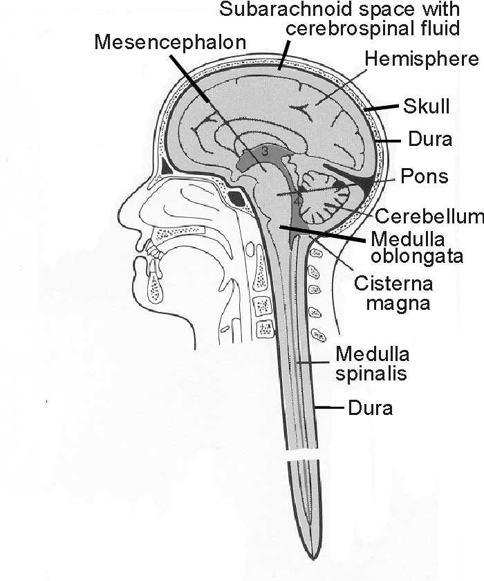
| Figure 3.1 The central nervous system seen in a
midsagittal section. |

|
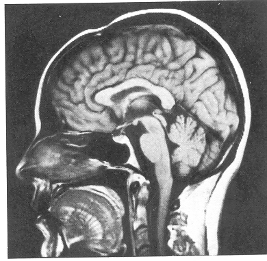
| Figure 3.2 Magnetic resonance image (MRI)
at a level corresponding to the drawing in Figure 3.1.
Most of the structures seen in Figure 3.1 can be
identified in this picture. Courtesy of Dr. S. J. Bakke,
The National Hospital of Norway, Oslo.
|
|
|
Same Anatomic Terms Used in This Book
The terms medial, toward the midline, and
lateral, away from the midline, are used to describe
the relative position of structures in relation to a
midsagittal plane of the body. The terms cranial or
rostral, toward the head or nose, and caudal,
toward the tail, are used to describe the relative
position of structures along a longitudinal axis of the body.
Thus, for example, nucleus A in the brain stem may lie medial
to and rostral to nucleus B, which in turn lies lateral to and
caudal to A. The terms ventral and dorsal
are used to describe the relative position of structures
in relation to the front (venter = belly) and the back
(dorsum) of the body, respectively. Anterior (front)
and posterior (rear) are most of ten used
interchangeably with ventral and dorsal (except for the human
forebrain, where anterior means toward the nose and ventral
means toward the base of the skull). |
the central nervous system. After some comments on the prenatal
development of the central nervous system, we start with a
description of the spinal cord, since conditions are simplest there.
THE SPINAL CORD
In humans the spinal cord is a 40 to 45 cm-long cylinder of
nervous tissue of approximately the same thickness as a little
finger. It extends from the lower end of the brain stem (at the
level of the upper end of the first cervica1 vertebra) down the vertebra1 cana1 (Figs. 3.1 and 3.3)
to the
=> page 73
start page
|