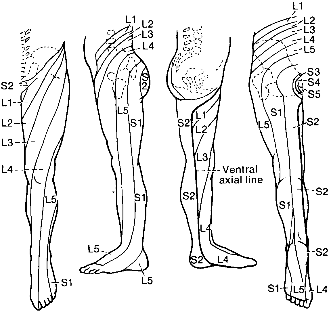
When a dorsal root is subjected to irriatation, as may happen by compression or stretching in connection with growth of an intraspinal tumor or protusion of an intervertebral disk, this causes pain and other sensory phenomena (numbness, pricking, tingling, and so forth) in the territory of the dermatome. Often the symptoms are felt only in smaller parts of the dermatome. With a protruding (herniated) intervertebral disk in the lumbar spine, for example, most often the roots of the fifth lumbar or first sacral nerves are affected (Fig. 7.13), and the pain is felt in the leg (sciatica).
How the dermatomes have been determined The dermatomal map presented here (Figs. 7.12 and 7.13, reproduced from Keegan and Garrett) is based on observations of a large number of patients with root compressions (usually due to a herniated intervertebral disk) and, in addition, on examination of the distribution of reduced sensation in volunteers subjected to local anesthesia of dorsal roots. The skin regions with reduced sensation (hypesthesia) were carefully mapped out before operation, and during operation it was determined which root was affected. Local anesthesia also produces a sensory loss much less extensive than the total distribution of sensory fibers of one dorsal root. Thus, the borders between dermatomes as presented in Figures 7.12 and 7.13 are imaginary. They ignore, for example, the great overlap between neighboring dermatomes, and on the other hand, that the dermatomes are much wider than the zones of hypesthesia occuring after damage to one dorsal root. It should be kept in mind that all dermatomal maps are composites of many single observations--in no single person have more than one or a few dermatomes been determined. For this reason, all maps showing dermatomes for the whole body can only be regarded as approximations, not taking into account, for example, the considerable individual variations that exist. This, together with the fact that different methods have been used, probably explains why the dermatomal maps of different authors vary so much. For the student the main emphasis should therefore be on learning certain main features of the dermatomal distribution rather than the artificial (and falsely accurate) borders indicated on the maps. |