|
Source:Understanding the nervous system An engineering perspective, 1993
by Sid Deutsch and Alice Deutsch
In the extracts below the author mentions that some people fear that, somehow, their minds may be read
from EEG signal recordings and then explains that this cannot be the case since the EEG signals
only reflect some kind of crude average of the many unresolved cortical potentials.
I believe this is a representative view among the wellinformed and this is why I
quoted it. Do note however, that the author in no way denies that by measuring those cortical potentials directly it becomes an entirely different mater.
There is also a passage stating that "..a dream remains the private property of its originator", which is credible in the context of EEG measurements, while this privacy does not extend to the context of direct connections to the nervous system.
The preface was quoted to give the reader an idea of what to expect, when actually consulting this source.
The title of the book is revealing and it is the kind of information an engineer, who has no access to classified information, will be looking for when contemplating the basic electronic aspects connected with cybernetic applications.
There is also a joking remark in the text advicing not to let a Ph D attach electrodes to a patient or anybody else.
Although it is stated jokingly I fear that in the context of these home pages,
it might be interpreted to mean something else. Since I am in favor of legitimate uses of all kinds of advanced technology for the benefit of mankind, I feel compelled to state my view that as long as there is a proper communication with the patient, there is no reason to block the use of any particular type of useful technology.
There is no reason to add to the irrational fears among the public.
What people should be warned about is the genuine danger of secret uses of technology for obscure purposes.
|
Preface
In 1987 a book that I wrote with Evangelia M-Tzanakou of Rutgers University, Neuroelectric Systems, was published by New York University Press. Sometime before this manuscript was completed it occurred to me that one ought to write a book, about the nervous system, for the lay person.
Of course, many books of this type have been written by life scientists, but a book written by a biomedical engineer would be different. Although some of my relatives and best friends are life scientists, there are two cultures. The world in general and the nervous system in particular look quite different to an engineer than they do to a life scientist.
I proceeded to generate a manuscript without equations (or, at least, nothing more complicated than Rate x Time = Distance). It was a challenge, a very enjoyable challenge, to explain the nervous system from an engineering perspective to the intelligent lay person. Alas, the manuscript was unpublishable. Even a literary agent did not help. Lay persons were undoubtedly interested in the nervous system, but not as seen through the eyes of an engineer.
Undaunted, I enlisted the aid of my daughter Alice, who is one of the life scientists that has a Ph.D. in biology. Alice made a semi-infinite number of changes, adding and subtracting so much material that it was only fair to call her a co-author. Unfortunately, this fooled nobody. The revised manuscript remained too heavily flavored by an engineering viewpoint.
Finally, I sent the manuscript to a publisher of engineering books - namely, IEEE PRESS. The PRESS's reviewers said, "Put the equations back, and you will have something that is viable!" I went even further-I cheerfully gathered up my favorite home work and exam questions, the residue of 25 years of teaching neuroelectric systems, and added Problems at the end of each chapter. (This includes answers to those exam gems that were formerly carefully guarded.)
One final change was made. One of the reviewers, Murray Eden of NIH, opined that most of the mathematics should be presented in appendix form at the end of each pertinent chapter. I am grateful to Dr. Eden for this excellent suggestion.
As the chapter headings indicate, the book begins with the basic components of the nervous system-with sensory receptors, neurons and their dendrites, and skeletal muscle circuits. This is followed by the more complicated auditory and visual systems. Finally, the climax - the brain - is considered. The 12 chapters correspond to a three-credit biomedical engineering course on the nervous system.
What is the engineering perspective? It is one in which each physiological system is simplified until it can be analyzed mathematically, followed by presentation as a simplified model.
The material in the book has been shaped by a myriad of students and colleagues-at the Polytechnic Institute of Brooklyn (now Polytechnic University) until 1972; at Rutgers University from 1972 to 1979; at Tel Aviv University from 1979 to 1983; and at the University of South Florida (as a "Visitor") since 1983. The manuscript was also modified in accordance with the helpful comments of some six "anonymous" reviewers, in addition to those of Dr. Eden.
I am thankful to the people of USF for their cooperation-especially Tom Smith and Dr. Elias Stefanakos of the Electrical Engineering Department, and Dr. Michael Kovac, Dean of Engineering. Special thanks are also due to the editorial staff of IEEE PRESS, notably Executive Editor Dudley Kay, Production Supervisor Denise Gannon, and Production Editor Karen Miller, without whose expertise this project could not have come to fruition.
Above all, Alice and I are grateful to Ruth, mother and wife, who was one of the "intelligent lay persons" who undertook to read the first few chapters of the first manuscript.
Sid Deutsch Sarasota, FL
Ever since I can remember, my father always answered my "why" and "how" questions about the world. No matter what he was doing, he always had time to give me a scientific explanation for some phenomenon of our environment. No question was ever answered by that oft-quoted phrase: "Look it up!" I still remember his description of Einstein's theory of relativity that we discussed when I was a preteen. When he came to me with the suggestion that I broaden his engineering interpretation of the nervous system, I was delighted. I saw this as my chance, in turn, to help him understand the whys and hows of the nervous system from the biologist's point of view. Biology relies so completely on experimentally proven facts. In contrast, I saw the book as a series of gross approximations about various aspects of the nervous system which had as starting points these facts. It was fascinating to me that in this way - using logical mathematical expressions and formulas - one could attempt to explain biological data. I suppose it is the dialectic of starting with some basic observations, developing a theory - in this case often a mathematical model - to explain the facts, and from this, designing better experiments so as to learn yet more facts which in turn will lead to a better mathematical description or model, and so on, until the "absolute" truth about the nervous system is known. It is my hope that this book will serve as a unique point of view about the nervous system that can stimulate both new research and new theories. I also hope that it can lead to a greater understanding of the nervous system by being valuable to both the bioengineer and the neurophysiologist.
Alice Deutsch New York, NY
- - -
1 - 3
(p 13)
...A "thought" that lasts for several seconds is the outcome of millions of cortical potentials chasing around inside the brain. A pair of electroencephalographic (EEG) electrodes attached to the head shows an unending stream of irregular discharges. It does not do much good to "think simple" during an EEG examination; the electrical signal remains incomprehensible. (Its magnitude is around 10
mV
1.)
[S. D. writes: "I once was approached by a patient who insisted that certain people were 'listening in' to his
inner thoughts by analyzing his EEG signals, and that they were able to do this without even using electrodes.
Alas, after many years of sophisticated monitoring of brain waves, one can only conclude that 'millions of
cortical potentials are chasing around inside the brain'; the nature of the signal radically changes during trauma
or epileptic seizures and during sleep, but a dream remains the private property of its originator."]
One final analogy may be helpful. Imagine that you are in a satellite looking down upon a large city. You cannot see individual people, of course. But if thousands of bodies march on the Capitol building, you may be able to see a blob, the integrated outcome of many individual images. Similarly, a pair of scalp electrodes can not "see" individual brain potentials on the other side of relatively thick skull bone. What it does see is the momentary movement of thousands of signals that happen to be "marching" in the same direction, but this direction changes many times in one second.
- - -
12-3 Electroencephalographic (EEG) Signals
In 1924, Hans Berger connected two electrodes (small, round, metal disks) to a patient's scalp and detected a feeble current by using a very delicate galvanometer. Berger thereby opened up a window on the brain. As electronic amplifiers and recording equipment came into common use, the brain signal - called an electroencephalogram (EEG) - became a major clinical tool of the neurologist [J. D. Bronzino, 1984; R. Elul, 1972; A. S. Gevins, 1984; A. S. Gevins et al., 1975; K. A. Kooi, R. P. Tucker & R. E. Marshall, 1978; M. Matonsek et al., 1967; G. Pfurtscheller and R. Cooper, 1975; A. Remond, 1971; E. Vaadia, H. Bergman & M. Abeles, 1989].
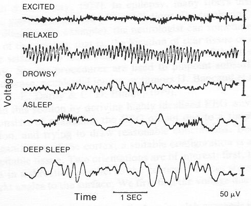
Five EEG records taken from a normal individual are shown in Fig. 12-4 [H. H. Jasper, 1941]. The time scale is given by the 1-s line at the bottom. The voltage scales are given by the 50-
mV
1 lines at the right side. For this type of recording, the "hot" electrode is usually located on the back of the head, opposite the striate cortex, and the other on a neutral point such as an ear lobe. In order to get a good recording, one must of course make good contact with the skin. It is not necessary to shave the patient's head, but skin oil should be removed with a solvent. A conducting paste is applied to the electrode, which is held by a suitable strap or cap against the scalp. The neutral ear-lobe electrode can be held in place with a simple clip.
[S.D. writes: "When I was at the Rutgers Medical School in New Jersey, I designed equipment for telemetering 15 simultaneous EEGs to a receiver in an adjoining room, via FM, so that the animal or human patient could be
|

| | Fig. 12-4 Electroencephalographic recordings taken from a normal individual. For this type of recording, the "hot" electrode is usually located on the back of the head, the other on a neutral point such as an ear lobe. In the "relaxed" waveform the patient's eyes are closed. Notice the time and voltage scales:
the 1-s line at the bottom and
50-mV 1 lines at the right side. (From Jasper, Epilepsy and cerebral localization, Thomas, 1941.)
free to move about. In the first test of the equipment I volunteered to be the patient. My neurologist friend and colleague insisted that the normal solvent treatment was inadequate for my oily skin, and proceeded to sandpaper my ear lobe until it bled. The recording was normal, but I was the buff of jokes for the next week or two about how my ear became bitten. The moral of the story-don't let a Ph.D. connect EEG electrodes to a patient or anybody else."]
The ear-lobe electrode does not pick up appreciable signals because it is relatively far away from electrical sources such as brain cells and muscle fibers; in fact, that is why it is considered to be a neutral point. The other electrode only "sees" the electrical activity in its immediate vicinity. To begin with, because of the thickness of scalp, muscles, skull bone, and mem- brane surrounding the brain, the electrode is around 1 cm away from the cerebral cortex. Signals from neurons deeper in the brain are rapidly attenuated as distance increases. The traces of Fig. 12-4 therefore reveal local happenings. The voltage from an individual neuron is far too small to show up on an electrode l cm away. What we are seeing are the voltages from thousands of neurons whose action or graded potentials happen to be headed in the same direction at the same time. It is somewhat like being up in a satellite and trying to see individuals on earth. If everybody in Times Square suddenly headed north, say, you could possibly see the flow in one direction. Normally, however, people moving one way cancel out the people moving in the opposite direction.
In the first waveform of Fig. 12-4, the patient is "excited" (but not agitated) in the sense that he (or she) is looking at the people in the room and listening to their conversations. To avoid interference from the APs that elicit muscle contraction, the patient is not talking or moving about. We see that the EEG is a weak
(10 mV 1 ), chaotic signal. From the frequencies present in the trace, we can conclude that it is not derived from high-speed (100 meters/second (m/sť APs, which would induce relatively high frequencies as they fly by. It is not even derived from slow 1 m/s APs. The frequencies are reminiscent of the 0.1-m/s progression of a graded potential of maximum amplitude 20 mV as it is conducted along a dendrite: its neuron integrates many inputs to generate its own graded potential, and the latter is "leisurely"
transmitted to another dendrite via an electrical gap junction or via a chemical agent that diffuses across a synaptic junction.
The fear voiced by some individuals that the neurologist can read their "inner thoughts" is without foundation.
Certain regularities can be observed if the person is looking at, say, a pattern of vertical stripes,
but the same thought repeated over and over again merely results in a random,
nonrepetitive and meaningless EEG pattern [Skeptical Jnquirer].
In the second waveform, which is "relaxed," the patient' s eyes are closed. This state gives rise to a relatively strong, nonsteady oscillation at around 10 Hz, known as the alpha rhythm [H. T. Castello, 1983; B. H. Jansen, 1985; J. G. Okyere, P. Y. Ktonas & J. S. Meyer, 1986]. The origin of the alpha rhythm is controversial, but it is probably connected to vision because it is strongest in the vicinity of the striate cortex. Especially mysterious is the fact that it vanishes when the patient opens his or her eyes and looks at the world about. A hypothetical explanation is offered at the end of this section.
As the patient progresses (or retrogresses) from relaxed to drowsy to asleep to deep sleep, radical changes in frequency and amplitude take place. "Deep sleep" displays slow waves, only a few cycles per second, of large amplitude
(100 mV 1). The large amplitude indicates that entire sections of tissue beneath the electrode are undergoing the same electrochemical changes in unison. The slow waves hint at the relatively slow transport of jons. Perhaps the EEG is telling us that the debris following many hours of wakefulness is being removed and "normal" material is being replenished. Altematively, in Chapter II, it is suggested that self-organization lakes place during "quiet" periods such as sleep. These are relatively slow processes; the human cycle consists of 16 hours of alertness followed by eight hours of sleep (not all of it, of course, as "deep sleep") [A. R. Morrison, 1983].
The clinical value of the EEG comes from the fact that an abnormal waveform is correlated with abnormalcy underneath the electrode [N. I. Bachen, 1986; D. G. Childers et al., 1982; C. D. McGillem, J. I. Aunon & K.-B. Vu, 1985; N. H. Morgan and A. S. Gevins, 1986; G. Pfurtscheller,
Footnote
1) microvolts.
Return to Introduction
Tillbaka till Introduktion
|
|
|